(800) 210-7017
PRP quality isn’t a single number stamped on a kit’s marketing slick, it’s a chain of variables that runs from the patient’s vein to the needle going into the tissue, and the kit only owns the middle of that chain. You’re judging the kit on platelet concentration, recovery efficiency, leukocyte profile, and red cell carryover, but you’re also judging the patient’s blood, the operator’s hands, and the anticoagulant and activator choices that bookend the spin.
A high-quality PRP is the product of matching kit chemistry to the clinical target and running the protocol with discipline, not just buying a premium kit.
There’s no single universal standard for PRP quality, which is why a handful of competing classification systems exist and why you have to read the labels carefully. In practice, clinics anchor quality to a small set of measurable parameters: platelet count per microliter, fold-increase over baseline, leukocyte profile with a neutrophil breakdown, and red cell contamination, all of which a standard complete blood count can report in one pass on the finished product.
| Classification | What It Reports | Best Use |
|---|---|---|
| DEPA | Dose, Efficiency, Purity, Activation | Full quality fingerprint |
| PAW | Platelet count, Activation, White cells | Quick clinical comparison |
| MARSPILL | Method, Activation, Spin, Light, Leukocyte content, and more | Research-grade detail |
One million platelets per microliter is commonly cited as the working clinical efficacy threshold, with most musculoskeletal targets running three to seven times whole-blood baseline.
The gap between what a kit’s brochure promises and what the kit actually delivers in your hands is one of the biggest sources of disappointment in PRP, and it’s where most buyers get burned. Concentration is also only half the story, because a kit can post an impressive multiple while quietly discarding most of the platelets through poor recovery efficiency, which means you need a larger draw to hit a useful absolute dose.
Chasing the highest platelet multiple isn’t automatically the right call, because growing evidence points to a U-shaped dose response where very high concentrations may inhibit healing in some tissues.
Separation method is the single biggest determinant of what your final product looks like, because it sets the upper and lower bounds on every other quality variable downstream. Pick the method to fit the clinical target: leukocyte-poor low-volume product from a single-spin system reads well for an arthritic knee, while a leukocyte-rich higher-concentration product from a double-spin earns its place in chronic tendinopathy work.
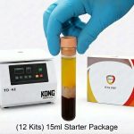
A single-spin gel system can be chairside-ready in under fifteen minutes, while a careful double-spin runs thirty or more, and that gap compounds across a busy injection day.
The leukocyte profile is one of the most clinically meaningful axes of PRP quality, and it cleanly splits products into two families with distinct use cases. The kit’s design largely picks the family for you: gel-separator single-spin kits produce leukocyte-poor output almost by default, while double-spin and buffy-coat kits produce leukocyte-rich output unless you deliberately discard the white cell layer.
| Profile | What’s Inside | Best Clinical Target |
|---|---|---|
| Leukocyte-poor (LP-PRP) | Most white and red cells stripped out | Intra-articular knees, hips, shoulders |
| Leukocyte-rich (LR-PRP) | Buffy coat retained with neutrophils, lymphocytes, monocytes | Chronic tendinopathy (lateral epicondylitis, patellar, Achilles) |
| Selective leukocyte | Lymphocytes and monocytes kept, neutrophils excluded | Newer preparations, hard to achieve reliably chairside |
Neutrophils release matrix-degrading enzymes that can damage cartilage intra-articularly, which is why a leukocyte-poor product is generally the right call for joints.
Residual red cells and neutrophils in the injectate are the under-discussed drivers of post-injection flare and inconsistent results, and they’re the contaminants you can actually do something about. Once injected, red cells lyse and release free hemoglobin, heme, and iron, which catalyze oxidative reactions that damage chondrocytes and tenocytes and produce the bruising-like pain patients report in the first twenty-four to forty-eight hours after a sloppy prep.
Even in a kit marketed as leukocyte-poor, a few percent of starting neutrophils can carry over and produce a disproportionate inflammatory response because they’re activated by handling.
Patient biology sets the ceiling for what any kit can produce, because the kit is only concentrating what arrives in the draw tube. A patient at 300,000 platelets per microliter walks in with nearly twice the raw material of one at 160,000, and no kit, no matter how good, manufactures platelets that aren’t there.
Patient prep, including a medication review and instructions to hydrate the day before, is part of producing high-quality PRP, not something separate from it.
The same kit run by two different clinicians can produce dramatically different products, and operator technique is the variable people underestimate the most. The venipuncture sets the quality ceiling before the centrifuge even spins, because a difficult draw with multiple sticks shears and pre-activates platelets, which means they release their growth factors into the tube instead of into the tissue where you need them.
Clinics that produce the most consistent PRP train one or two designated staff extensively and run periodic complete blood counts on finished product as a quality audit.
A high platelet count means little if the platelets show up at the injection site already spent, so kits and protocols are judged by whether they keep platelets viable and releasable until they hit the target tissue. Premature activation in the tube is the primary failure mode, triggered by rough handling, excessive g-force, contact with non-siliconized surfaces, air exposure at the meniscus, or under-anticoagulation, and when it happens the injectate becomes a dilute supernatant of growth factors instead of a loaded reservoir.
A growth factor ELISA ratio comparing baseline release in unactivated product to burst release after calcium-chloride or thrombin activation is a more honest viability measure than total growth factor content alone.
The variables that operate in the last few minutes before injection are easy to overlook, but they can decide whether a well-prepared PRP behaves the way you intended in the tissue. Anticoagulant choice, activation strategy, and elapsed time from draw to needle all stack on top of everything the kit and centrifuge already locked in, and they’re where careful protocols separate themselves from casual ones.
Many orthopedic protocols deliberately skip pre-activation and rely on endogenous activation when PRP contacts native collagen and thrombin at the lesion site, producing a slower physiologic release pattern over hours rather than seconds.